At The Brain and Spine Centre, we specialize in the diagnosis and surgical treatment of neurological and spinal disorders. Dr. Muhammad Aqeel Natt, a leading neurosurgeon in Lahore, provides advanced, safe, and compassionate care for patients.
Brain Tumours Biopsy
Brain tumor biopsy requires expert precision and diagnostic accuracy to guide optimal treatment decisions. At The Brain and Spine Centre, Dr. Muhammad Aqeel Natt provides specialized brain biopsy services at Farooq Hospital, West Wood Branch, Lahore, combining advanced stereotactic techniques with compassionate support.
Our goal is simple: obtain accurate tissue diagnosis safely, guide treatment strategy, and support informed decision-making for the best possible outcomes.
What Is a Brain Tumour Biopsy?
A brain tumor biopsy is a diagnostic procedure involving removal of a small tissue sample from an abnormal area of the brain for examination under a microscope. The tissue sample is analyzed by a pathologist to determine the presence, type, and grade of tumor, infection, inflammation, or other disease processes. This information is essential for recommending appropriate treatment and predicting prognosis.
Types of brain biopsy include:
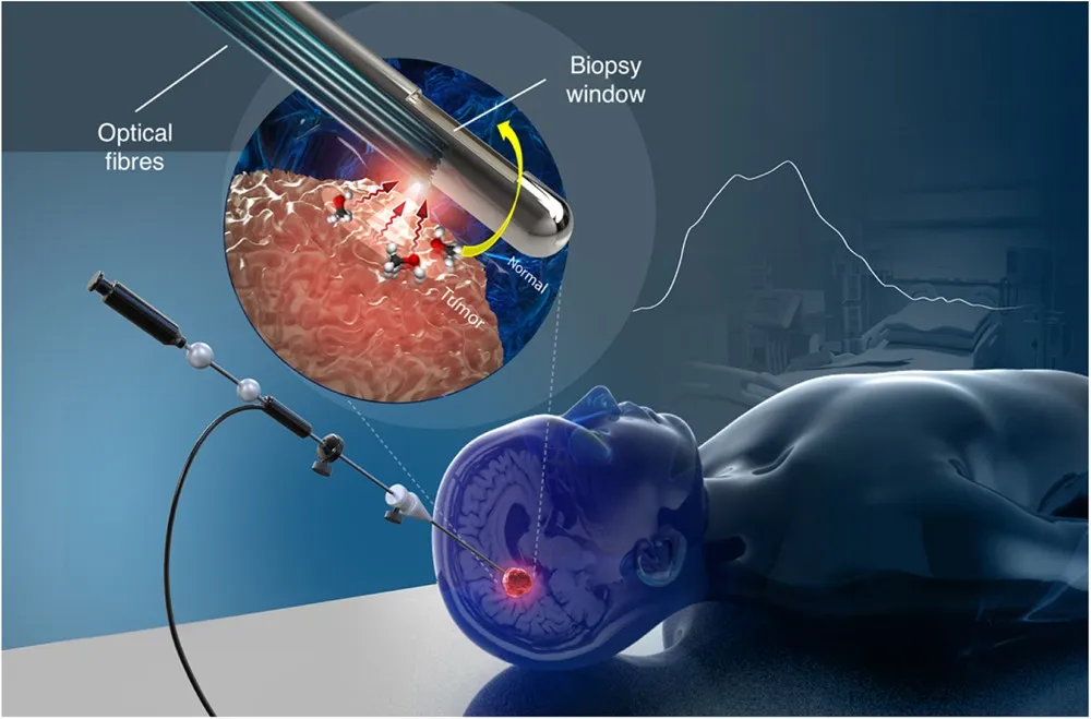
Stereotactic needle biopsy is a minimally invasive procedure using advanced imaging (MRI or CT) and three-dimensional computer navigation to precisely guide a biopsy needle to the target lesion through a small burr hole. This is the most common method and is preferred for deep-seated tumors and lesions in eloquent brain areas.
Open biopsy involves removing part of the skull (craniotomy) to obtain a larger tissue sample and is used when there is higher risk of bleeding or when a larger specimen is needed.
Neuroendoscopic biopsy uses an endoscope to access intraventricular and paraventricular lesions through minimally invasive approaches.
Biopsy during tumor removal may be performed as part of the surgery to remove all or part of the tumor.
Symptoms Requiring Brain Tumour Biopsy
Brain biopsy is typically recommended when imaging studies reveal lesions of uncertain cause that require tissue diagnosis before treatment can begin.
Common indications for brain biopsy include:
Multifocal or diffusely infiltrating tumors not suitable for debulking surgery, deep-seated tumors not amenable to safe surgical resection, tumors in eloquent brain areas where resection would cause significant neurological deficits, elderly patients harboring large tumors either multi-centric or deep-seated, radiological suspicion of primary CNS lymphoma requiring tissue confirmation before initiating treatment, multiple brain lesions where systemic cancer is not confirmed, and lesions that cannot be distinguished from infection, inflammation, or radiation necrosis on imaging alone.
Diagnosis
The biopsy procedure itself establishes the diagnosis by providing tissue for pathological examination.
Pre-biopsy imaging:
MRI is the preferred imaging modality, providing detailed soft tissue images to identify the target lesion, plan the safest trajectory, and avoid critical structures. MRI may include spectroscopy to assess chemical composition of the tumor.
CT scan is used with the stereotactic frame in place to calculate precise coordinates for needle placement.
Pathological examination by a neuropathologist determines tumor type, grade, and molecular characteristics essential for treatment planning. Preliminary frozen section analysis may be performed during the procedure to confirm adequate tissue sampling, with final results available in 5-7 days.
Treatment Options
Brain biopsy is a diagnostic procedure, not a treatment. However, the information obtained guides treatment decisions.
Stereotactic needle biopsy procedure:
A stereotactic frame or frameless navigation system is applied to the patient’s head. Imaging is performed to localize the target and calculate coordinates. A small scalp incision (approximately 1 cm) is made and a burr hole (dime-sized) is drilled in the skull. The biopsy needle is guided precisely to the target lesion using real-time navigation. Multiple small tissue samples (typically 4 samples) are removed for examination. The needle is withdrawn and the incision is closed with sutures. The entire procedure typically takes one hour.
Diagnostic yield:
Brain biopsy has a diagnostic yield of 89-97% for brain tumors. For non-neoplastic conditions, diagnostic yield ranges from 57-72%. The procedure successfully identifies alternative diagnoses in approximately 35% of suspected neuroinflammatory disorders.
Therapeutic impact:
Brain biopsy changes treatment strategy in approximately 77% of patients by confirming diagnosis, guiding therapy selection, or allowing safe initiation of treatment by excluding alternative diagnoses.
The Procedure
Our process ensures safety, precision, and diagnostic accuracy:
Consultation: Comprehensive evaluation including review of imaging studies, discussion of biopsy indication, risks, benefits, and alternatives.
Pre-operative imaging: MRI under neuronavigation protocol to plan the safest biopsy trajectory.
Stereotactic biopsy: Frame or frameless navigation applied, followed by contrast-enhanced CT for coordinate calculation. Minimally invasive needle biopsy performed under local anesthesia with sedation or general anesthesia.
Pathological examination: Tissue samples sent immediately to neuropathologist for preliminary and final analysis.
Post-procedure monitoring: CT scan performed after biopsy to check for complications, followed by observation for at least 4 hours.
Recovery & Aftercare
Recovery from stereotactic brain biopsy is typically quick with minimal downtime. Most patients require only an overnight hospital stay, with many able to go home the same day. Hospital observation typically lasts 1-2 days for monitoring. Most people resume normal activities within one to two days.
Post-procedure care includes:
Neurological assessments performed in the recovery room and during hospitalization to monitor for complications. Post-operative CT scan to detect hemorrhage or other complications. Pain management with prescribed medications for sensitivity or headache around the incision site. Monitoring for symptoms requiring immediate attention including severe headache, worsening neurological deficits, seizures, or fever.
Biopsy results timeline:
Preliminary frozen section results may be available within one hour during the procedure. Final pathology results typically take 5-7 days, though complex cases may require up to 10 days. Your doctor will discuss results and treatment options at a follow-up appointment.
Results You Can Expect
High diagnostic accuracy with diagnostic yield of 95-97% for brain tumors and 62-72% overall for unexplained CNS disorders. Repeat biopsy increases diagnostic yield when initial biopsy is non-diagnostic.
Safe procedure with low complication rates. Symptomatic complications occur in only 4.5% of cases. Serious complications causing permanent neurological deficit or death occur in approximately 1-1.5% of patients.
Therapeutic impact in 77% of patients by guiding treatment decisions, allowing appropriate therapy initiation, or avoiding unnecessary treatments.
Definitive diagnosis enabling selection of optimal treatment including surgery, radiation, chemotherapy, immunotherapy, or observation based on tumor type and grade.
Why Choose The Brain and Spine Centre
Led by Dr. Muhammad Aqeel Natt, specialist neurosurgeon with extensive expertise in stereotactic brain biopsy and minimally invasive neurosurgical techniques. Access to advanced imaging technologies including high-field MRI and CT for precise biopsy planning and real-time navigation. State-of-the-art stereotactic equipment for accurate targeting with minimal risk to surrounding brain tissue. Collaboration with expert neuropathologists for rapid preliminary diagnosis and comprehensive final pathology reports. Multidisciplinary tumor board approach to discuss biopsy results and recommend optimal treatment strategies. Convenient location at Farooq Hospital, West Wood Branch, Lahore with modern operating suite and recovery facilities.
Cost of Brain Tumour Treatment
Costs vary with biopsy technique, imaging requirements, anesthesia type, duration of hospitalization, and pathology testing complexity. Personalized estimates provided after consultation and imaging review.
Frequently Asked Questions (FAQs)
Can I know Dr. Muhammad Aqeel Natt’s credentials?
What types of brain tumours do you treat?
Is the surgery safe?
Do I need long-term follow-up after surgery?
Do I need long-term follow-up after surgery?
Are you having health problems? Contact us today!
Address Business
Contact With Us
Call Us 24/7: 0318 4065914
Working Time
Sunday: 8.30am - 19.30pm