At The Brain and Spine Centre, we specialize in the diagnosis and surgical treatment of neurological and spinal disorders. Dr. Muhammad Aqeel Natt, a leading neurosurgeon in Lahore, provides advanced, safe, and compassionate care for patients.
Percutaneous Transpedicular Fixation
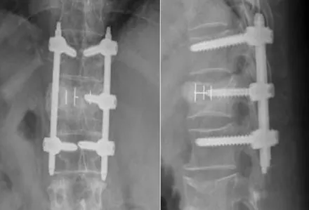
Percutaneous transpedicular fixation (PTPF) is a minimally invasive surgical technique for spinal stabilization that achieves the same biomechanical fusion and fixation as traditional open posterior instrumentation while dramatically reducing soft tissue trauma, blood loss, and recovery time. At The Brain and Spine Centre, Dr. Muhammad Aqeel Natt offers advanced percutaneous transpedicular fusion at Farooq Hospital, West Wood Branch, Lahore, using image-guided fluoroscopic and navigation techniques for precise screw placement and early patient mobilization.
Our goal is simple: stabilize the unstable spine, promote fusion, and allow rapid return to function with minimal surgical morbidity.
What Is Percutaneous Transpedicular Fixation?
Percutaneous transpedicular fixation is a minimally invasive approach to spinal stabilization in which pedicle screws are placed through small percutaneous incisions (typically 8–12 mm per side) using fluoroscopic or neuronavigational guidance, then connected with rods passed through subcutaneous tunnels to create a rigid, load-sharing construct. Unlike traditional open posterior fusion, which requires large paravertebral muscle dissection, PTPF minimises soft tissue injury while achieving equivalent mechanical stability.
Indications for PTPF
PTPF is suitable for a broad range of spinal conditions requiring stabilization and fusion.
Common indications:
Degenerative lumbar spine disease: spondylolisthesis (grade I–II), recurrent or persistent discogenic back pain despite conservative therapy.
Lumbar spinal stenosis with instability: when decompression alone is insufficient and adjacent segment degeneration is a concern.
Herniated disc disease: particularly recurrent herniated discs at the same level.
Spinal trauma/fractures: unstable thoracolumbar and lumbar fractures, particularly in polytrauma patients where minimising surgical morbidity is beneficial.
Spinal deformity: selected cases of adult scoliosis or kyphosis, where less invasive stabilisation is preferred.
Post-operative instability or revision: adjacent-segment degeneration after previous fusion; previous laminectomy creating instability.
Spinal metastases and tumours: pathologic fractures and instability from cancer.
Symptoms and Patient Selection
Candidates typically present with:
Chronic back pain, radicular symptoms, or leg pain despite 6–12 weeks of conservative treatment (physical therapy, anti-inflammatory drugs, injections).
Progressive neurological deficits or instability on imaging.
Overall good health without major contraindications to surgery; morbidly obese patients or those with severe medical comorbidities may have higher complication risk.
Diagnosis and Preoperative Planning
Detailed imaging and assessment guide surgical planning.
X-rays (standing or dynamic): show degree of listhesis, vertebral alignment, prior fusion levels, and bone quality.
CT scan: high-resolution imaging of pedicle anatomy, vertebral body, and fracture patterns; essential for measuring pedicle diameter and defining entry points.
MRI: evaluates disc herniation, stenosis, cord compression, and assesses need for concurrent decompression.
Sagittal alignment analysis: determines degree of correction and instrumentation strategy.
The Procedure
PTPF is performed under general anaesthesia with fluoroscopic or neuronavigational guidance.
Key surgical steps:
Positioning: prone on operating table with abdomen free to reduce venous pressure and bleeding.
Percutaneous screw placement:
Small skin incisions (8–12 mm each) placed over planned pedicle entry points.
Using AP and lateral fluoroscopic views, a cannulated needle or trocar is advanced into the pedicle (upper outer quadrant) under real-time imaging guidance.
A guidewire is placed, and the track is tapped; a cannulated screw is then inserted over the wire.
In experienced hands, fluoroscopy-guided or navigation-assisted placement ensures accurate screw positioning with minimal cortical perforation.
Rod passage and connection:
After bilateral screw placement, a rod (or rods) is carefully tunneled through subcutaneous tissue to connect the screw heads.
Rods are secured to screws with set screws and locking mechanisms, creating a rigid construct.
Decompression (if needed): concurrent lumbar decompression (laminotomy, facetectomy, discectomy) can be performed through separate small stab incisions or a mini-open approach without sacrificing the benefits of minimal-access instrumentation.
Recovery & Aftercare
Recovery is typically much faster than open surgery.
Hospital stay: 1–3 days for uncomplicated cases; most patients walk on postoperative day 1.
Pain and mobility: significantly less postoperative pain, earlier mobilisation, and faster return to activities compared to open surgery.
Fusion progression: bone-to-bone contact and load sharing promote fusion; most patients achieve solid fusion at 6 months.
Return to work/activity: light duties may resume within 2–4 weeks; full activity within 2–3 months as healing progresses.
Physiotherapy: postoperative exercises strengthen core muscles and support spinal stabilisation.
Results You Can Expect
With appropriate patient selection and technique:
Pain relief: significant improvement in back pain (VAS and Oswestry Disability Index scores) in >90% of cases.
Functional outcome: most patients achieve good-to-excellent functional recovery with early mobilisation and return to work.
Fusion rates: solid fusion achieved in >95% of cases at 6–12 months when combined with intervertebral grafting (interbody fusion).
Complication profile: lower rates of infection, blood loss, and soft tissue-related complications compared to open fusion; screw malposition risk is minimised with fluoroscopy/navigation guidance.
Revision safety: if revision is needed, percutaneous instrumentation can often be removed with minimal morbidity, allowing restoration of spinal mobility.
Why Choose The Brain and Spine Centre
Led by Dr. Muhammad Aqeel Natt, with extensive expertise in minimally invasive spine surgery, percutaneous pedicle screw placement, and image-guided techniques. Access to advanced fluoroscopic and neuronavigational imaging to ensure precise screw placement, avoiding neurovascular and visceral complications. Capability to combine percutaneous fixation with decompression, interbody fusion (MI-TLIF, MI-ALIF), and correction of deformity in a single, minimally traumatic procedure. Emphasis on early mobilisation and return-to-function protocols, with dedicated physiotherapy support.
Cost of Percutaneous Transpedicular Fixation
Costs depend on number of levels, need for concurrent decompression or interbody fusion, imaging guidance complexity, and hospital stay length. PTPF may reduce overall cost and morbidity compared to traditional open fusion due to shorter ICU stay and faster recovery. Detailed cost estimates are provided after imaging review and surgical planning.
Frequently Asked Questions (FAQs)
Can I know Dr. Muhammad Aqeel Natt’s credentials?
What types of brain tumours do you treat?
Is the surgery safe?
Do I need long-term follow-up after surgery?
Are you having health problems? Contact us today!
Address Business
Contact With Us
Call Us 24/7: 0318 4065914
Working Time
Sunday: 8.30am - 19.30pm