At The Brain and Spine Centre, we specialize in the diagnosis and surgical treatment of neurological and spinal disorders. Dr. Muhammad Aqeel Natt, a leading neurosurgeon in Lahore, provides advanced, safe, and compassionate care for patients.
Stereotactic Biopsy
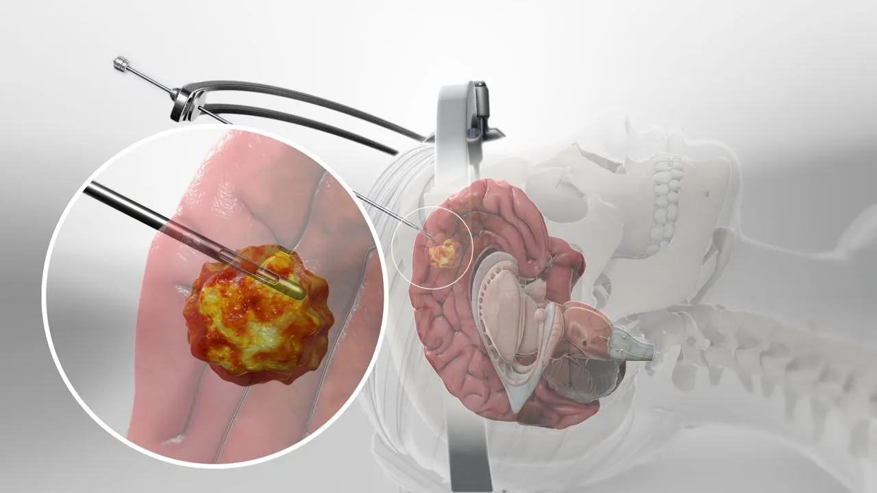
Stereotactic biopsy is a minimally invasive, highly accurate diagnostic procedure that obtains tissue samples from brain lesions of uncertain cause using precise three-dimensional imaging guidance. At The Brain and Spine Centre, Dr. Muhammad Aqeel Natt performs stereotactic brain biopsy at Farooq Hospital, West Wood Branch, Lahore, using modern frame-based and frameless techniques with excellent diagnostic accuracy and minimal patient morbidity.
Our goal is simple: obtain accurate tissue diagnosis safely, guide treatment decisions, and minimize brain trauma.
What Is a Stereotactic Biopsy?
Stereotactic biopsy is a surgical technique that uses advanced imaging (MRI or CT) combined with three-dimensional computer guidance to precisely localize and sample abnormal brain tissue through a small needle or hollow probe, without requiring full surgical exposure of the lesion. The term “stereotactic” refers to the three-dimensional coordinate system used to map the brain and target specific lesions with millimeter accuracy. Stereotactic biopsy is particularly valuable for deep-seated, multifocal, or poorly accessible lesions where open surgery carries unacceptable risk.
Two main approaches:
- Frame-based stereotaxy: Uses a rigid stereotactic frame attached to the skull for precise target definition; typically performed under local anesthesia.
- Frameless (neuronavigation) stereotaxy: Uses surface fiducials and intraoperative tracking; offers greater patient comfort and flexibility, though may require general anesthesia.
Symptoms and Indications
Stereotactic biopsy is recommended when imaging reveals an unexplained brain lesion requiring tissue diagnosis before treatment can begin.
Common indications include:
- Multifocal lesions suggesting lymphoma, infection, or metastatic disease.
- Deep-seated lesions in the basal ganglia, thalamus, brainstem, or ventricles inaccessible to safer open biopsy.
- Lesions in eloquent brain areas (motor, language, visual) where open surgery risks severe disability.
- Suspected infection (tuberculoma, fungal infection, toxoplasmosis) requiring organism identification.
- Suspected primary CNS lymphoma, which requires tissue diagnosis before chemotherapy.
- Differentiating radiation necrosis from recurrent tumor.
Diagnosis
Pre-biopsy evaluation identifies the target and determines technical approach.
- Clinical history and neurological exam establish baseline function and any neurological deficits.
- High-resolution MRI (T1, T2, FLAIR, DWI sequences) localizes the lesion, identifies the safest biopsy trajectory avoiding eloquent areas and vascular structures, and assesses internal heterogeneity to guide optimal sampling sites.
- CT scan may complement MRI by showing calcifications or bony landmarks.
- Vascular imaging (MR or CT angiography) identifies major vessels to avoid during needle passage.
The Procedure
Stereotactic biopsy is performed under general or local anesthesia depending on technique.
Frame-based approach:
- Stereotactic frame is applied to the skull under local anesthesia.
- Thin-cut CT or MRI is obtained with fiducials visible; frame coordinates are calculated based on lesion location and safe trajectory.
- A burr hole is created at the calculated entry point.
- Biopsy needle is advanced under frame guidance to the target lesion.
- Multiple small tissue samples (typically 4–6) are obtained, with intraoperative frozen-section analysis confirming adequate tissue sampling.
Frameless approach:
- Fiducials (tracking markers) are placed on the scalp under local anesthesia or on the head after induction of general anesthesia.
- Preoperative MRI or CT is used for image registration and target planning.
- Intraoperative neuronavigation system tracks the biopsy probe in real-time relative to the preoperative images.
- Biopsy needle is advanced to target under imaging guidance.
- Tissue samples are obtained; frozen-section analysis confirms adequacy.
Recovery & Aftercare
Stereotactic biopsy is minimally invasive with rapid recovery.
- Hospital stay: 1–2 days or same-day discharge in selected cases.
- Postoperative imaging: CT or MRI performed after biopsy to exclude hemorrhage or other complications.
- Diagnostic results: Preliminary results from frozen-section analysis may be available during surgery; final pathology typically takes 5–7 days.
- Activity resumption: Most patients resume normal activities within 1–2 days, with minimal incision care.
Results You Can Expect
Diagnostic accuracy:
- Overall diagnostic yield ranges from 89.4% to 98.5% depending on careful technique and on-demand frozen-section analysis.
- Both frame-based and frameless techniques achieve comparable accuracy with no significant differences.
- Non-diagnostic or inconclusive results occur in 1.5–10.6% of cases.
Safety profile:
- Procedure-related morbidity is low (1–13.3%), with new or worsened neurological deficits in only 1–13.3% of cases.
- Infections occur in <7% of cases.
- Procedure-related mortality is extremely rare (0%).
- Seizures occur in 10% but are usually manageable.
Clinical impact:
- Accurate tissue diagnosis changes management in >75% of patients, guiding chemotherapy, radiation, immunotherapy, or supportive care.
- 60% of patients experienced improved functional status following diagnosis and appropriate treatment.
Why Choose The Brain and Spine Centre
Led by Dr. Muhammad Aqeel Natt, specialist neurosurgeon with extensive experience in stereotactic brain biopsy using both frame-based and frameless techniques. High-resolution MRI and CT with neuronavigation planning to ensure precise targeting and safe needle trajectories. On-demand intraoperative frozen-section analysis dramatically improves diagnostic yield and ensures adequate tissue sampling. Expert neuropathology consultation for rapid, accurate diagnosis and optimal treatment recommendations. Safe, minimally invasive approach with low morbidity and high patient satisfaction.
Cost of Stereotactic Biopsy
Costs vary with imaging requirements, frame or frameless technique, hospital stay, intraoperative frozen-section analysis, and pathology testing complexity. Personalized cost estimates provided after consultation and imaging review.
Frequently Asked Questions (FAQs)
Can I know Dr. Muhammad Aqeel Natt’s credentials?
What types of brain tumours do you treat?
Is the surgery safe?
Do I need long-term follow-up after surgery?
Are you having health problems? Contact us today!
Address Business
Contact With Us
Call Us 24/7: 0318 4065914
Working Time
Sunday: 8.30am - 19.30pm